
Figure 3.
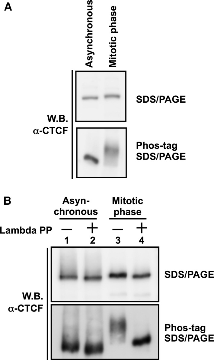
Phosphorylation of mitotic CTCF. (A) Analysis by phos‐tag SDS/PAGE. Whole cell lysates were separated on a 7.5% SDS/PAGE (upper panel) and a 5% SDS/PAGE containing 20 μm phos‐tag (lower panel), followed by western blotting with anti‐CTCF antibody. (B) Lambda phosphatase treatment of mitotic CTCF. Cell lysates prepared from asynchronous cells (lane 1) or mitotic‐arrested cells (lane 2) were subjected to immunoprecipitation assays using anti‐CTCF antibody. The immunoprecipitated CTCF was incubated in the absence or presence of lambda protein phosphatase and subjected to phos‐tag SDS/PAGE as described above.