
Figure 1.
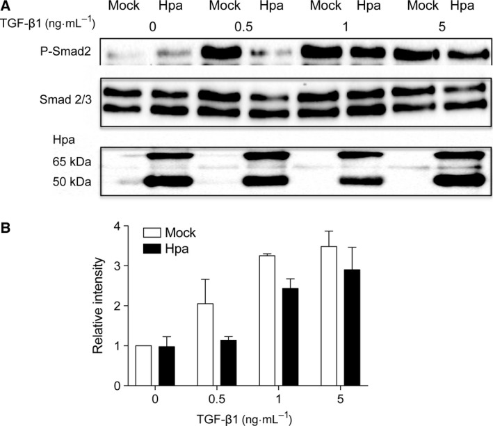
TGF‐β1‐induced Smad phosphorylation in Mock vs. Hpa Fadu cells—Fadu cells stably overexpressing human heparanase (Hpa) and mock (Mock) transfected cells were seeded into six‐well plates at a density of 6 × 10⁵ cells per well. After 24 h of starvation (serum‐free medium), the cells were stimulated for 30 min with TGF‐β1 at the indicated concentrations. (A) Lysate supernatants were analyzed by western blotting using anti‐phospho‐Smad2 and anti‐Smad2/3 antibodies. Overexpression of heparanase in the Hpa cells was confirmed using anti‐heparanase antibody. (B) Band intensity measured in three independent experiments was analyzed by image lab™ Software and the average band intensity of P‐Smad2 is shown. Band intensity value of Mock cells without TGF‐β1 stimulation is defined as 1.