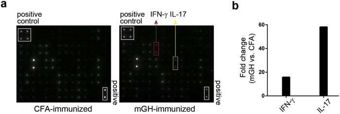
Figure 2. Heightened productions of IFN-γ and IL-17 by pituitary-infiltrating cells in mouse autoimmune hypophysitis.
Single-cell suspensions from the pituitaries of CFA-immunized mice or mGH-immunized mice were stimulated by mGH. Cytokine productions after stimulation were detected by cytokine array membranes. The dots that represent IFN-γ and IL-17 on the membrane are indicated (a). Signal intensity of IFN-γ and IL-17 was measured for the two groups of mice. Differences in the cytokine level, expressed as fold change (mGH vs. CFA), were derived by dividing the signal intensity of mGH group by the signal intensity of the CFA group (b).

