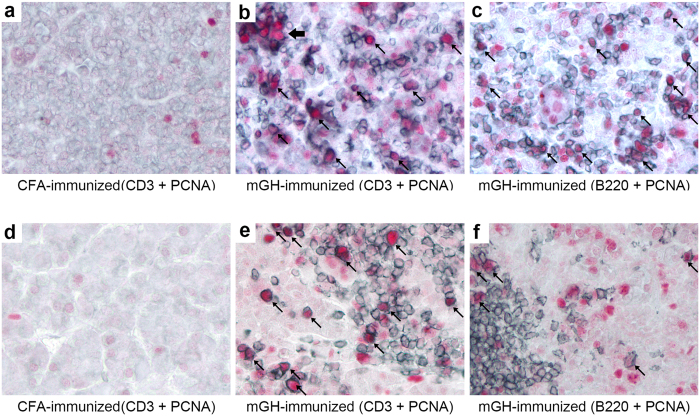
Figure 4. Persistent T and B cell proliferation in the pituitary gland of mouse autoimmune hypophysitis.
Pituitary sections of mice that develop autoimmune hypophysitis on day 28 (b and c) or day 56 (e and f) were immunostained by PCNA (in red) and CD3 (b and e, in blue) or B220 (c and f, in blue). Proliferating cells are indicated by arrows. Note a small cluster of T cells that were stained by PCNA (thick arrow) in (b). Pituitary sections from CFA-immunized mice were immunostained for PCNA and CD3 as a negative control (a and d).

