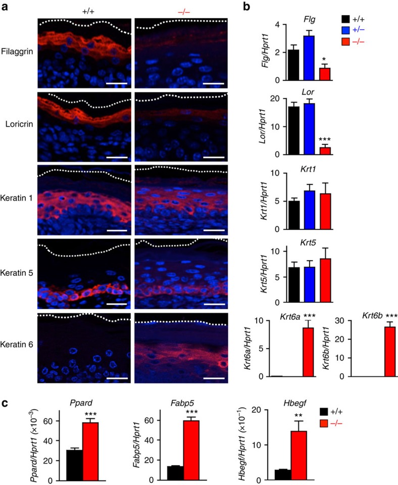
Figure 2. Aberrant terminal differentiation of Pnpla1−/− epidermis.
(a) Immunohistochemical staining of keratinocyte differentiation markers (red), followed by conterstaining with DAPI (blue), in skin sections from Pnpla1+/+ and Pnpla1−/− newborn mice. Scale bars, 20 μm. (b) qPCR analysis of keratinocyte differentiation markers in newborn Pnpla1+/+, Pnpla1+/− and Pnpla1−/− epidermis (n=5 animals per group). (c) qPCR analysis of PPARδ (Ppard) and its potential target genes in newborn Pnpla1+/+ and Pnpla1−/− epidermis (n=7 animals). In b,c, values are mean±s.e.m.; *P<0.05, **P<0.01, and ***P<0.001 versus Pnpla+/+ mice. Representative results from two or three independent experiments are shown.

