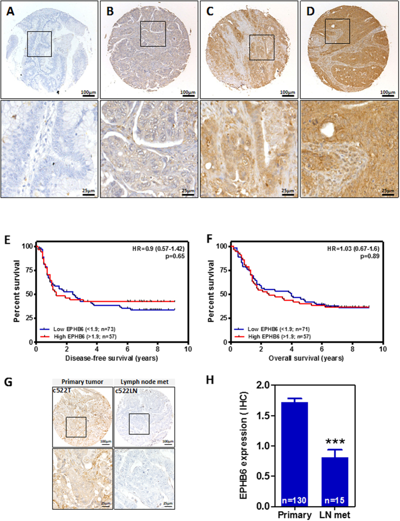
Figure 7. EPHB6 expression in primary colorectal tumors and patient survival.
(A,D) The levels of EPHB6 protein expression were determined by immunohistochemistry using sections of a tissue microarray containing formalin-fixed, paraffin-embedded tumor samples from a cohort of 130 Dukes C colorectal cancer patients. Representative images of colorectal tumors with different levels of EPHB6 are shown in panels (A,D). No associations were observed between EPHB6 expression and disease-free (E) or overall (F) survival of these patients. The Logrank test p values are shown. (G) The levels of EPHB6 protein expression in a representative primary tumor and a matched lymph node metastasis is shown. (H) Histogram showing the average (±SEM) EPHB6 immunointensity score in primary colorectal tumors and lymph node metastasis (Student’s t-test p < 0.0001).

