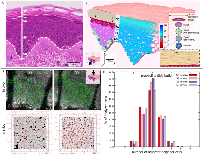
Figure 1. The biomechanical model of ellipsoid cells allows to faithfully recreate epidermal morphology.
(A) (I) Using ellipsoid cells, we can attain realistic tissue morphology as observed in histological preparations, including the typical epidermal layering consisting of stratum basale (SB), stratum spinosum (SS), stratum granulosum (SG), and stratum corneum (SC). (II) Our biomechanical model leads to the typical interdigitating SC pattern (inset). The left half of the 3D tissue cross-section shows colouring according to cell differentiation status. The right half displays cellular water content. Inner blue circles represent cell centres (not shown for SC due to the narrow cell size). (B) (I) Topological analysis of SG and SS based on manually segmented cells in in vivo images and cross-sectional images of the in silico epidermis. (II) The probability distribution of nearest neighbours per cell in silico follows closely the in vivo distribution.

