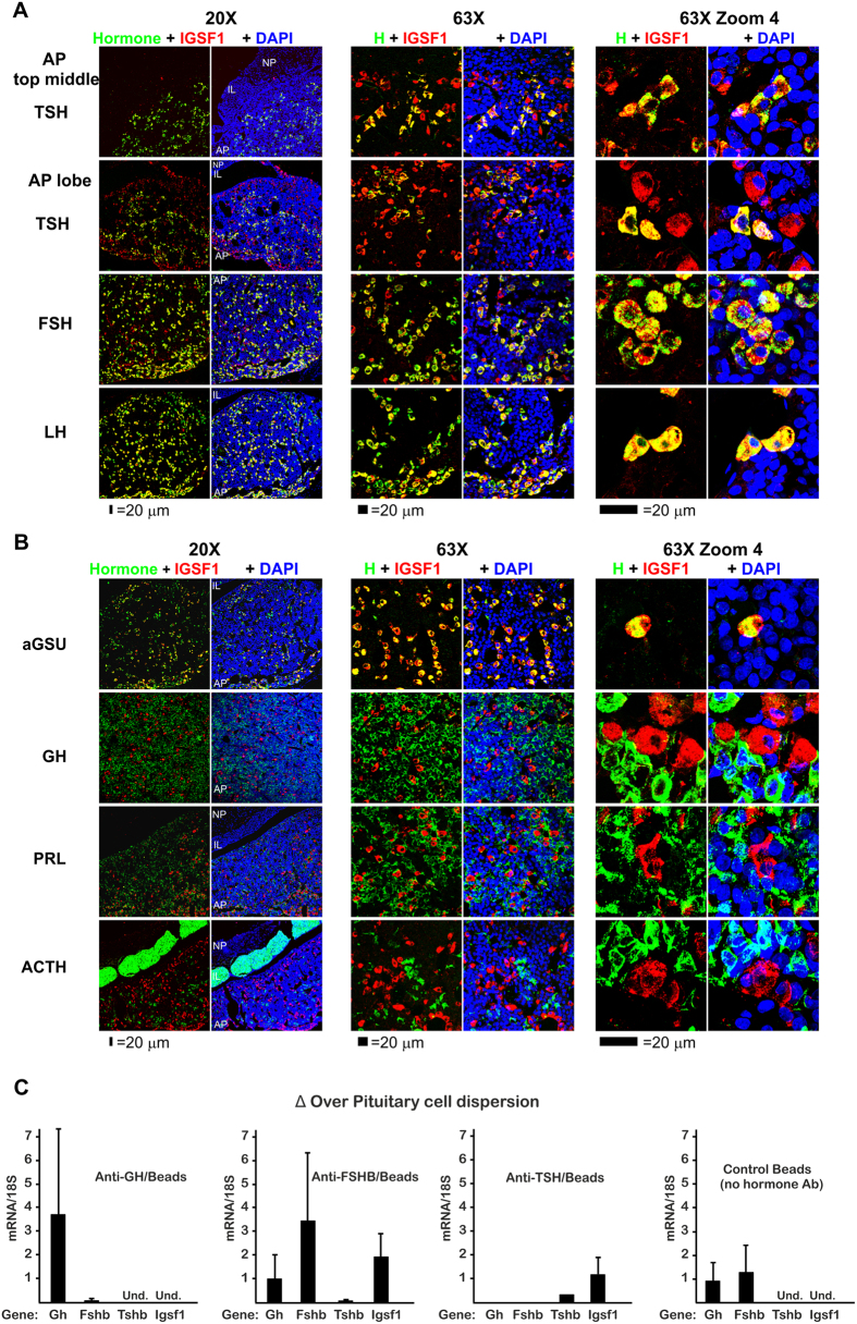
Figure 2. IGSF1 cellular expression in young adult male rat pituitary gland is expressed in thyrotrope and gonadotrope endocrine cells.
Coronal pituitary sections were stained and topographical serially studied using confocal microscopy from the middle towards the lobes at different magnifications (20X, 63X and 63XZoom4). A white laser was used to prevent differences in intensity by use of different wavelength lasers. IGSF1 is shown in red pseudocolor while every hormone is shown in green. Nuclei were stained with DAPI and shown in blue. (A) IGSF1 is located exclusively in the AP but not in the IL or the NP and co-localizes with the three hormones: TSH beta, FSH beta and LH beta. The number of double positive IGSF1/TSH was higher in the middle of the section (thyrotrope region) than toward the lobes (gonadotrope region). (B) All IGSF1 cells co-localize with aGSU, the common alpha subunit for TSH, FSH and LH. No somatotrope (GH), lactotrope (PRL) or corticotrope (ACTH) was found to be expressing IGSF1. Quantifications are shown in Supplemental Fig. 2B. (C) Immune-magnetic purification of somatotropes (anti-GH/Beads), gonadotropes (anti-FSHB/Beads) and thyrotropes (anti-TSHB/Beads) from a single cell dispersion of rat pituitary followed by qRT-PCR. Results are expressed as enrichment (gene mRNA/18 S) over the initial cell dispersion. As a control, magnetic beads in the absence of hormone antibody were used. As expected, Gh mRNA was enriched in anti-GH, Fshb mRNA in anti-FSHB and Tshb mRNA in anti-TSHB purified cells. Igsf1 mRNA was detected exclusively in the anti-FSHB and anti-TSHB purified cells. Results are the mean of four independent experiments. AP = adenopituitary. IL = intermediate lobe. NP = neuropituitary.

