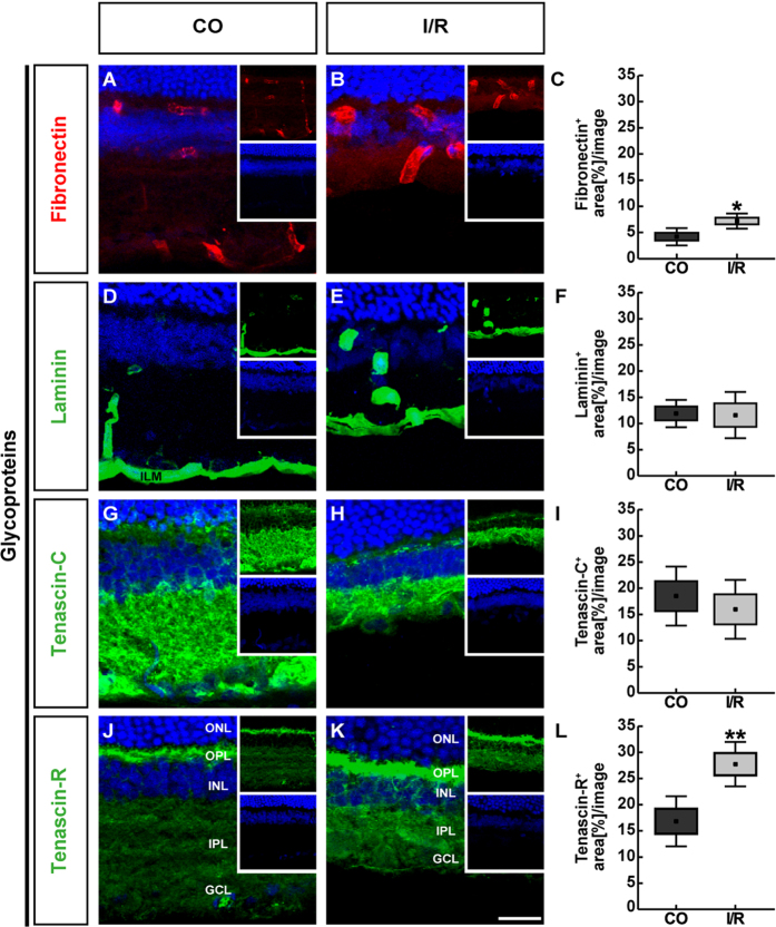
Figure 2.
Representative retinal cross-sections of control (CO) and ischemic (I/R) eyes stained using specific antibodies directed against the ECM glycoproteins fibronectin (A,B, red), laminin (D,E, green), tenascin-C (G,H, green) and tenascin-R (J,K, green) as well as the nuclear dye TO-PRO-3 (blue) shown as merged images. Small inserts display TO-PRO-3 (blue) and glycoprotein (green/red) staining separately. In both experimental groups, fibronectin as well as laminin displayed a blood vessel-associated staining. In addition, laminin was found in the ILM and in the GCL. Tenascin-C and -R staining was mainly localized in the IPL, OPL and GCL. Quantification revealed a significant increase in the fibronectin and tenascin-R staining area in ischemic retinae, whereas no significant changes were observed regarding the tenascin-C and laminin staining (C,F,I,L). Values are mean ± SEM ± SD. *p ≤ 0.05; **p ≤ 0.01; n = 5/group. GCL = ganglion cell layer, ILM = inner limiting membrane, INL = inner nuclear layer, IPL = inner plexiform layer, ONL = outer nuclear layer, OPL = outer plexiform layer. Scale bar = 50 μm.

