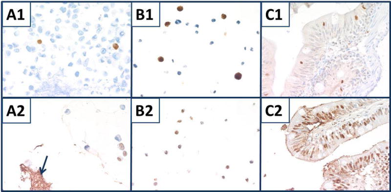
Fig. 10.

Staining for apoptosis. Top panels were stained for cleaved caspase 3. A1) Nine combined cell lines with splenic stroma. B1) SU-DHL-6. C1) Mosquitofish intestine. Bottom panels were stained with TUNEL assay. A2) Nine combined cell lines with splenic stroma (blue arrow). B2) SU-DHL-4. C2) Mosquitofish bowel. All panels, original magnification × 400.
