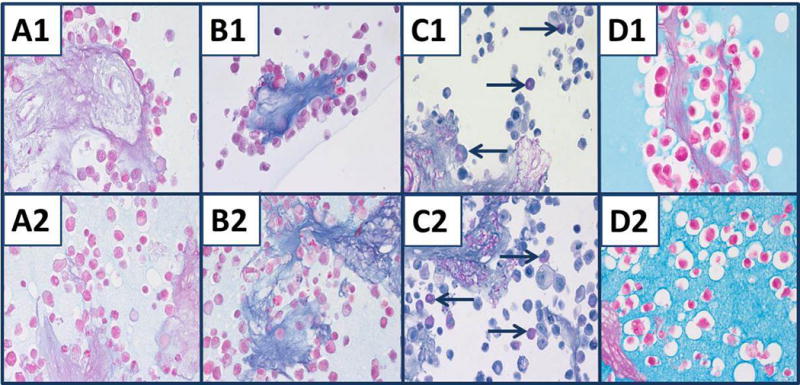
Fig. 2.

Mucin staining of nine combined cell lines and acellular stroma of spleen. Cell lines and stroma embedded in either HistoGel™ (#1) or 1.2% agar (#2). A1, A2) AB-1.0. B1, B2) AB-2.5 stains stroma of spleen blue, but there is little staining of cells other than nuclear counterstain. C1, C2) Selective cells stained with PASH and PAS of splenic stroma (blue arrows). D1, D2) Colloidal iron does not stain splenic stroma clearly, but does stain the HistoGel™ matrix (D1) and agar matrix (D2). Staining of cells and splenic stroma by colloidal iron is not apparent owing to staining of the matrices, HistoGel™ or 1.2% agar. All panels, original magnification × 400.
