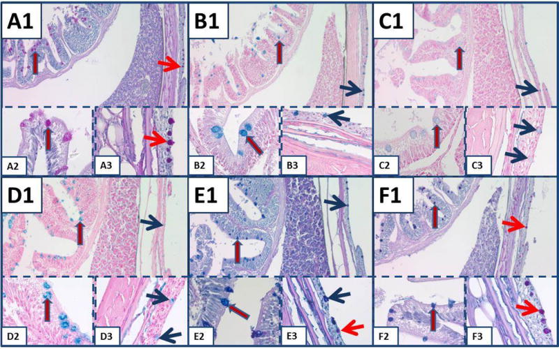
Fig. 3.

Mucin staining in the small intestine and skin of mosquitofish. A) PASH stains goblet cells of small intestine (red arrow blue outline) and goblet cells of the skin (red arrow). Similar arrows are used for the same identifications in subsequent panels, but colors demonstrating the goblet cells of the skin are red or blue to differentiate staining. B) AB-2.5. C) AB-1.0. D) Colloidal iron. E) AB-2.5-PASH. In panel E3, note the different types of mucin in the goblet cells of the skin. F) Colloidal iron-PASH. In (F), different types of mucin are stained in goblet cells of skin (red) compared to goblet cells of intestine (blue). All panels, original magnification × 100; insets × 630.
