
Fig. 1.
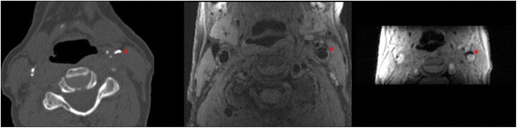
Example of calcification in the left carotid artery bifurcation (indicated by the red star) on CT (left image) and on CMR (middle image; PDw-FSE-BB sequence, and right image; magnitude image of the 3D-phase contrast sequence)
Official websites use .gov
A
.gov website belongs to an official
government organization in the United States.
Secure .gov websites use HTTPS
A lock (
) or https:// means you've safely
connected to the .gov website. Share sensitive
information only on official, secure websites.
Example of calcification in the left carotid artery bifurcation (indicated by the red star) on CT (left image) and on CMR (middle image; PDw-FSE-BB sequence, and right image; magnitude image of the 3D-phase contrast sequence)