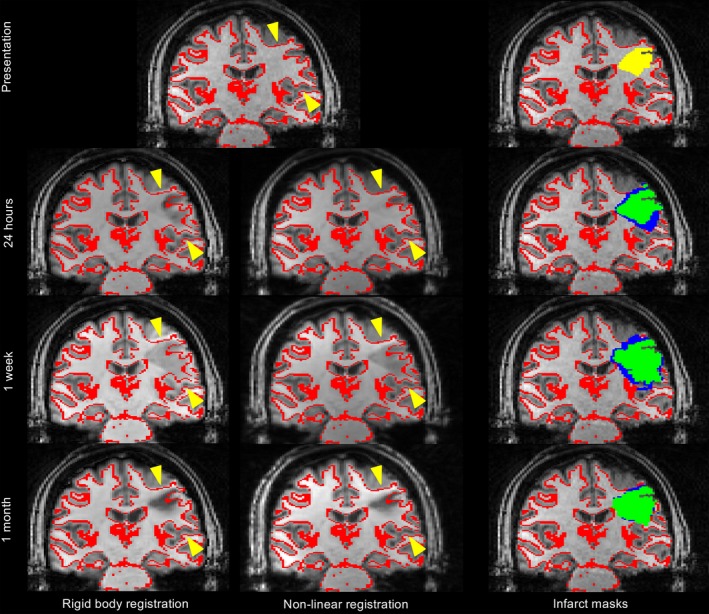
Figure 2.

Rigid body versus nonlinear registration in a patient presenting with right‐sided weakness and sensory loss. The top row shows the presenting T1‐weighted structural image with the gray‐white matter interface (red) overlaid for reference, and with the presenting ADC infarct overlaid (yellow, top right image). The lower rows show the T1‐weighted structural images from each follow‐up timepoint (24 h, 1 week, and 1 month) registered to the presenting scan using either rigid body or nonlinear registration, with the presenting gray‐white matter interface overlaid for reference (red). Yellow arrows highlight the regions where rigid body registration does not correct for subacute edema at 24 h and 1 week. The right hand column shows the infarct masks from each time point overlaid on the presenting T1‐weighted structural image using either rigid body (blue) or nonlinear registration (green).
