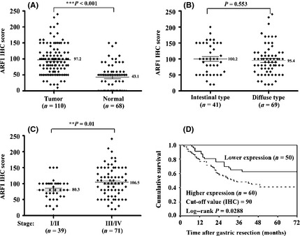
Figure 3.

Immunohistochemical (IHC) detection of ADP‐ribosylation factor 1 (ARF1) expression in human gastric cancer tissues (T) and matching non‐cancerous mucosa (N). (A) Comparison of the ARF1 IHC scores in T/N. The differences were analyzed with the Wilcoxon signed rank test. Comparison of the ARF1 expression in two types (B) or various stages (C) of cancer by the Mann–Whitney U‐test. (D) Kaplan–Meier survival curves of two groups of gastric cancer patients defined by the ARF1 expression level cut‐off value of 90, as determined with IHC scoring. The 5‐year survival rate of the lower expression group (n = 50) was significantly better than that of the higher expression group (n = 60; P = 0.0288, log–rank test).
