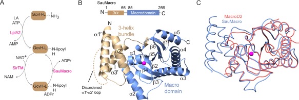
Figure 1.

Overall structure of SauMacro. A. Lipoate/ADPr cycle. B. Overall fold of SauMacro. SauMacro is rendered as ribbons with secondary structures indicated, where amino‐terminal helices α1′‐α3′ are colored tan, carboxy‐terminal macrodomain fold is colored blue, and Zn2+ is rendered as a magenta sphere. C. Cα trace overlay of SauMacro (blue) with human MacroD2 (red). Structures were aligned using MatchMaker in Chimera.26 SauMacro Zn2+ is rendered as a sphere (magenta). All figures were prepared in Chimera26 unless otherwise indicated.
