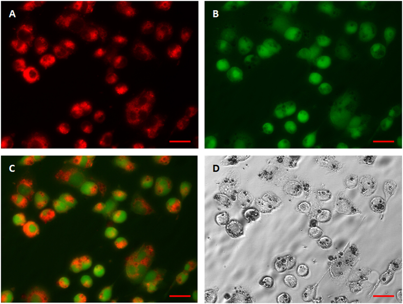
Figure 7. Fluorescent microscopy images of the MCF-7 cellular uptake of DiI-labelled microparticles and their intracellular distribution.
(A) Under 549 nm laser excitation, red fluorescence represents microparticles; (B) Under 488 nm laser excitation, green fluorescence represents cells; (C) Merged picture of (A,B); (D) Brightfield image. After incubation with DiI-labelled microparticles for 4 h, many microparticles had been phagocytized by the cells. Scale bars represent 20 μm.

