
Figure 7. Effects of Nr1d1-silencing on the cell cycle.
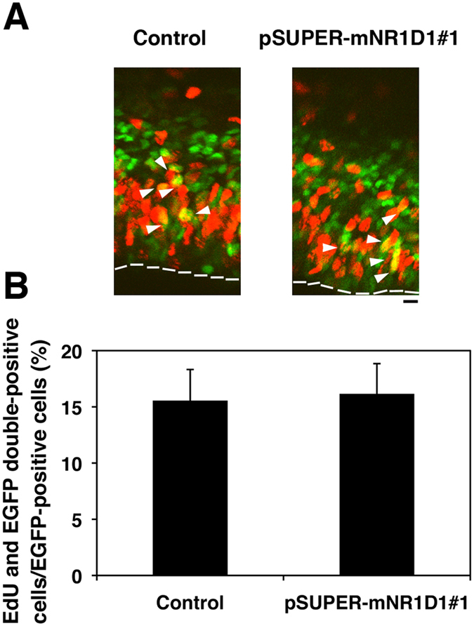
(A) E14.5 cortices were coelectroporated with pCAG-GFP together with control pSUPER vector or pSUPER-mNR1D1#1. The subsequent procedure performed is described in “Materials and methods”. Coronal sections were immunostained for GFP (green) and EdU (red). Dotted lines represent ventricular surface. Bar, 10 μm. (B) Quantification of EdU/GFP double-positive cells relative to GFP-positive ones in (A). Error bars indicate SD (n = 3).