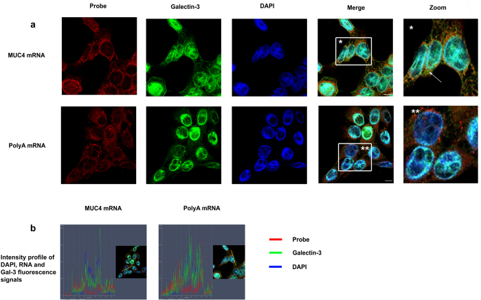
Figure 6. Galectin-3 interacts with mature mRNAs in the cytosol.
(a) PolyA mRNA or MUC4 specific transcripts were detected by FISH using either a Cy3.5 oligodT 40 mer or a Cy3.5 MUC4 specific 48 mer probe directed against the tandem repeat (red) in Sc cells. Galectin-3 was detected by IF (green). Nuclei were stained by DAPI (blue). Analyses were performed using laser scanning confocal microscopy and the pictures show a single z-plane. Co-localization was assessed by merging the images (Merge) where yellow spots indicate co-localization of transcripts and Gal-3. A zoom is performed in the area delimitated by a white square and arrow highlights example of co-localization. Scale bar: 10 μm. (b) Fluorescence intensity profiles of Gal-3, DAPI and RNA probe staining in Sc cells. Fluorescence intensity was quantified across the cell along the yellow arrow (see inset) using the ZEN software (Zeiss microscope) and presented as a histogram (blue, DAPI; green, Gal-3; red, RNA probe). The profile displayed is representative of three carried out independently.

