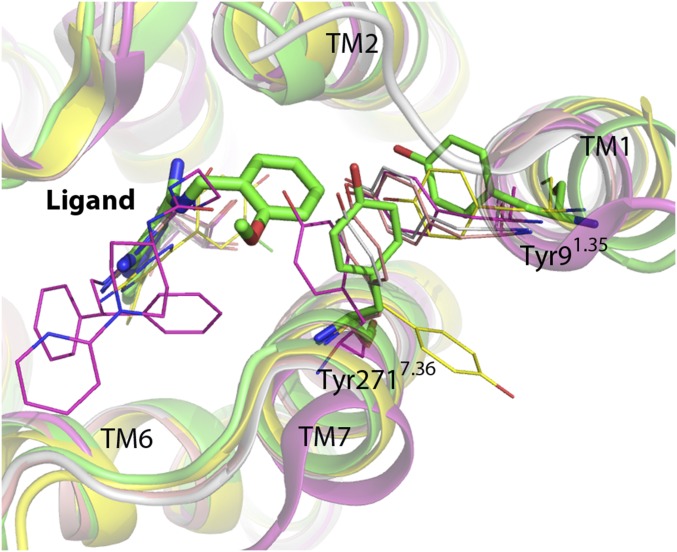
Fig. S5.
Comparison of Tyr91.35 and Tyr2717.36 positions and rotamers with other A2AR structures. The structure of A2AR–BRIL–Cmpd-1 (green) is superimposed with A2AR-T4L–UK-432097 (magenta; PDB ID code 3QAK), A2AR-TS–adenosine (salmon; PDB ID code 2YDO), A2AR-TS–NECA (salmon; PDB ID code 2YDV) and A2ARTS–T4E (yellow; PDB ID code 3UZC). Tyrosine side chains and ligands are shown in stick for A2AR–BRIL–Cmpd-1 and in lines for the other structures.

