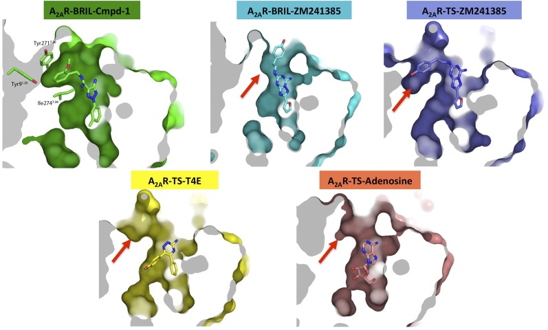
Fig. S7.
Comparison of the allosteric pocket in A2AR–BRIL–Cmpd-1 structure to equivalent parts in other reported A2AR structures. The structures shown here include A2AR–BRIL–ZM241385 (cyan; PDB ID code 4EIY), A2AR-TS–ZM241385 (blue; PDB ID code 3PWH), A2AR-TS–1,2,4-triazine antagonist (A2AR-TS-–T4E, yellow; PDB ID code 3UZC), and A2AR-TS–adenosine (salmon; PDB ID code 2YDO). These structures are all aligned with the A2AR–BRIL–Cmpd-1 structure (green), and surfaces are all viewed and clipped at same position for comparison. Red arrows point to the equivalent part in each structure and the sites are either very shallow pockets or clefts exposed to solvent.

