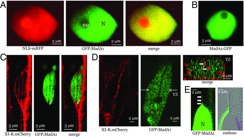
Fig. 3.
Analysis of GFP:MadA1 localization relative to nucleus-specific marker NLS-mRFP (fused to NLS derived from SV40 T-antigen) or XI-K:mCherry (myosin XI-K tagged by mRFP Cherry). (A) Colocalization of NLS-mRFP and GFP:MadA1. (B) Nuclear localization of MadA1:GFP. (C and D) Colocalization of XI-K:mCherry and GFP:MadA1. In D, the nuclear cross-section along the y–z plane marked by white arrows (Center) is shown (Upper Right); white arrows point to red, XI-K:mCherry-tagged material present within the nucleus. (E) GFP:MadA1-containing material apparently exported from the nucleus (N) as a linear array of discrete bodies marked by white arrows.

