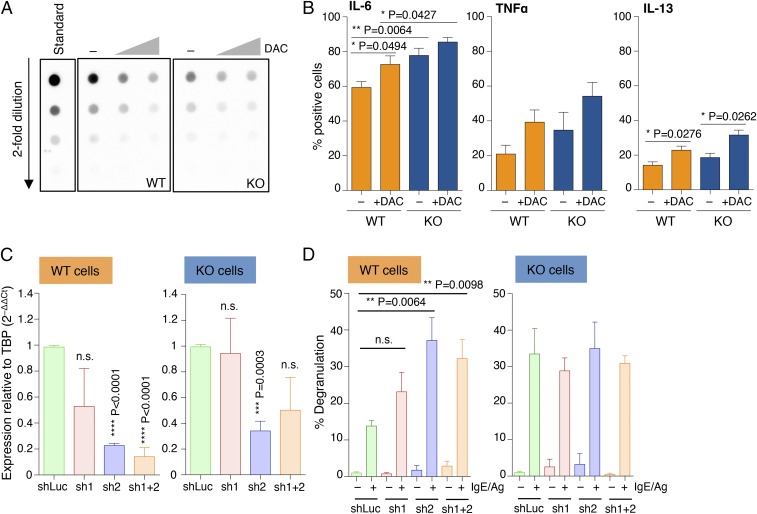
Fig. 3.
Increased mast cell responses upon disruption of DNA methylation activities. (A) Differentiated mast cells were treated with 0.5 and 5 μM 5-aza-2′-deoxycytidine for 48 h, after which genomic DNA was extracted and overall levels of 5mC were quantified by dot blot. (B) Mast cells were treated with 0.5 μM DAC for 3 d before stimulation with IgE and antigen and measurement of cytokine production by intracellular cytokine staining. Mean ± SEM; unpaired t test, two-tailed. (C) Mast cells were transduced with lentiviral vectors (sh1 and sh2) expressing shRNAs to knock down expression of Dnmt1. A vector expressing an irrelevant hairpin (shLuc) was used as control. After puromycin selection of the transduced cells, total RNA was extracted and the extent of Dnmt1 down-regulation was measured by qRT-PCR. Shown are the compiled results of three independent experiments. Mean ± SEM; unpaired t test (relative to the shLuc sample), two-tailed. (D) Same as C, except that cells were stimulated with IgE and antigen prior to measurement of the extent of degranulation by β-hexosaminidase release. Shown are the compiled results of three independent experiments. Mean ± SEM; unpaired t test, two-tailed.

