Fig. 4.
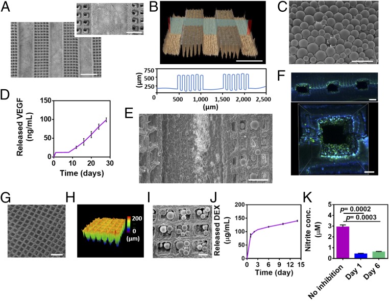
Fabrication of a predefined vascular layer and sustained release system layers. (A) SEM micrographs of micropatterned tunnels and the cage-like structures between them. (B) Confocal image of the microtunnels (Upper) and a topography analysis (Lower). (C) SEM micrograph of PLGA microparticles. (D) Cumulative release of VEGF. (E) SEM micrographs of PLGA microparticles deposited within the cage-like structures adjacent to the microtunnels. (F) Immunofluorescent images of CD31 (green) in endothelial cells cultured within the microtunnels to form lumens. Cell nuclei are shown in blue. (G and H) SEM (G) and confocal (H) micrographs of micropatterned cage-like structures. (I) SEM micrograph of PLGA particles deposited on cage-like structures. (J) Cumulative release of DEX. (K) Anti-inflammatory activity of DEX released from PLGA microparticles as indicated by the inhibition of NO secretion (measured as nitrite, a stable metabolite of NO) from activated macrophages. The results represent mean values ± SEM (n ≥ 5 in each group). Statistical evaluations were performed by unpaired Student’s t tests. (Scale bars: 500 μm in A and B; 200 μm in A, Inset, F, Upper, and G; 100 μm in C, E, and F, Lower; 50 μm in I.)

