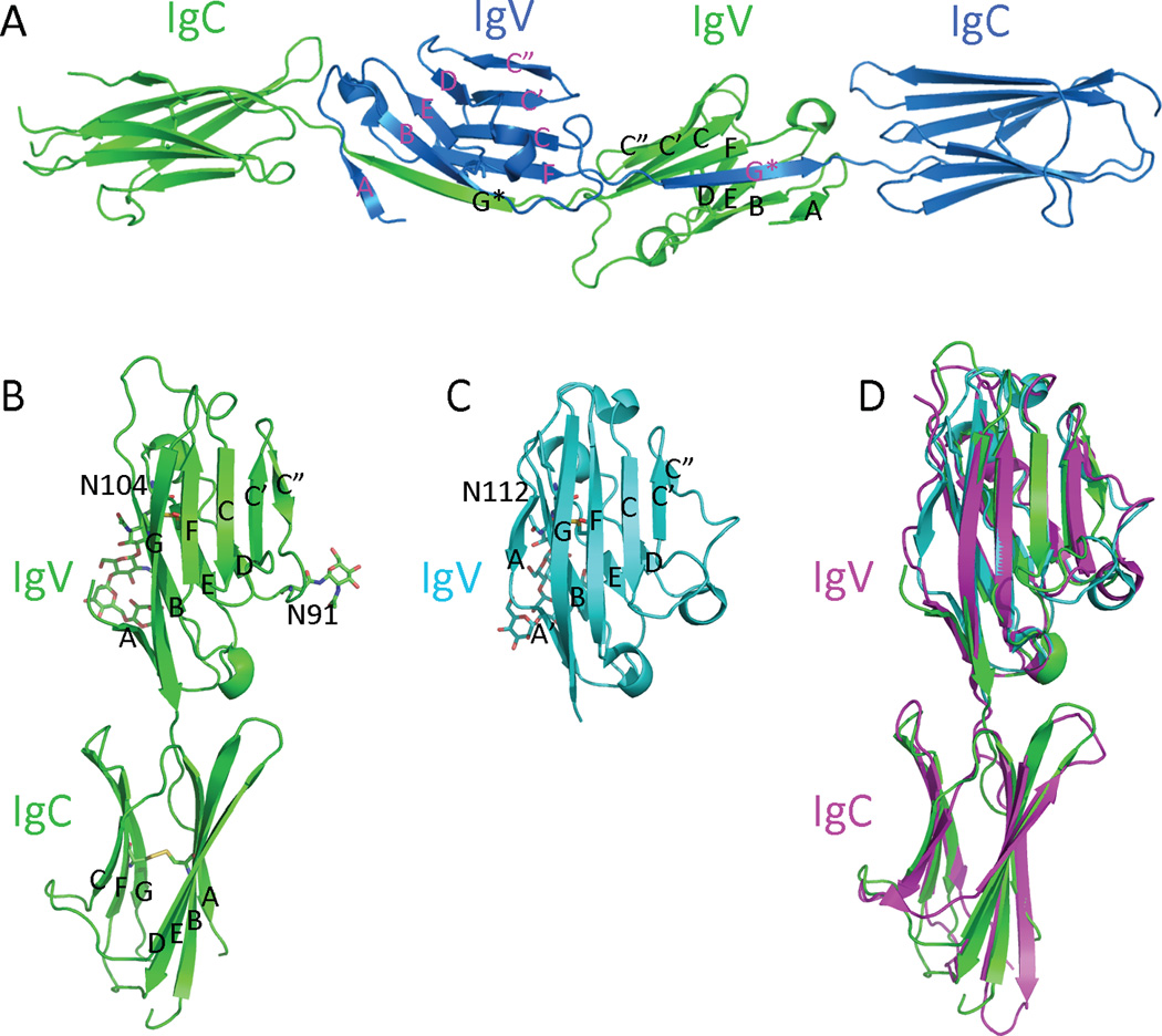
Figure 1. Structures of B7-H3, B7x and PD-L1.

(A) Overall structure of the dimeric mB7H3 in the crystal (PDB entry 4I0K). The strands from each monomeric mB7H3 are colored and labeled differently. (B) The generated model of monomeric mB7H3 based on the crystal structure from PDB entry 4I0K. (C) The structure of the hB7x IgV domain (PDB entry 4GOS). (D) Superimposition of the monomeric mB7H3, hB7x and hPD-L1 (PD-L1 is from A chain of the PDB entry 3BIK). The disulfide bonds, carbohydrates and the connecting Asn residues are shown as sticks.
