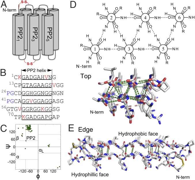
Fig. 1.
sfAFP structure (PDB ID code 2PNE). (A) Connectivity with the disulfide bonds is noted in red. (B) Sequence with helical regions (underlined), valines and lysines (red), and the two PG turns (blue). (C) Ramachandran map of native structure with four major basins noted. (D) Top and (E) edge view highlighting the internal H-bond network and the flat hydrophilic and hydrophobic faces.

