Fig. 1.
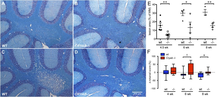
CRYAB expression determines severity of cuprizone-induced demyelinating pathology. (A–D) LFB staining of myelin in cerebellar tissue of control mice (A and B) or mice treated with cuprizone for 8 wk (C and D) shows that cuprizone-induced demyelination is more severe in WT mice (A and C) than in Cryab−/− mice (B and D). (E) Quantification of cerebellar lesion size, as percentage of white matter area, in WT (closed symbols) and Cryab−/− mice (open symbols) after 4.5, 6, and 8 wk of cuprizone diet, reveals that at all time points lesions in WT mice are significantly larger than in Cryab−/− mice. Shown are data points representing individuals animals, mean ± SEM, and P values determined by unpaired t test (n = 4; *P ≤ 0.05, **P < 0.01). (F) Motor skill performance, as measured by a rotarod assay and depicted as percentage change from baseline performance, reveals that WT mice (blue boxes) are more susceptible to cuprizone-induced motor skill deficits than Cryab−/− mice (red boxes). Data are shown as box-and-whisker plots, with whiskers depicting minimal and maximal values. P values were determined by unpaired t test (n = 8–16; †P < 0.1, *P < 0.05).

