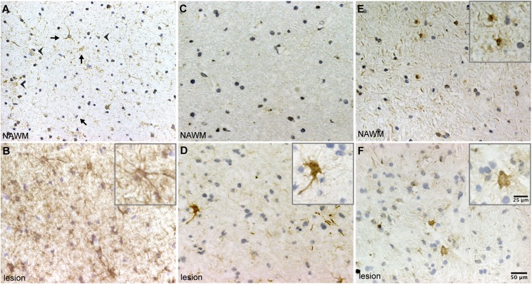
Fig. 4.
CRYAB in astrocytes in MS lesions is preferentially phosphorylated at serine-59. (A and B) Immunohistochemical analysis of CRYAB in NAWM (A) and areas with active demyelination (B) demonstrates that CRYAB is expressed by astrocytes (arrows) and oligodendrocytes (arrowheads) in NAWM (A), and that expression of CRYAB is increased in astrocytes with reactive morphology in active demyelinating areas (B). (C–F) Analysis of CRYAB, phosphorylated at Ser-59 (C and D) and Ser-45 (E and F), in NAWM (C and E) and active demyelinating areas in MS tissue (D and F), reveals that phosphorylation of Ser-59 is not present in NAWM (C) and is dramatically increased in cells with reactive astrocyte morphology in active demyelinating lesions (D), whereas phosphorylation of Ser-45 is present in both NAWM (E) and active demyelinating areas (F). Nuclei were counterstained with hematoxylin. Insets depict magnified images of positive-staining astrocytes.

