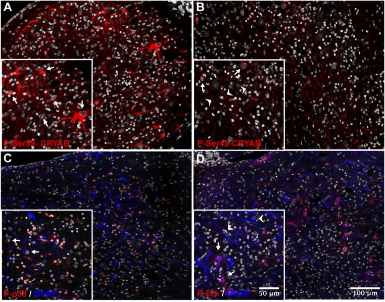
Fig. 5.
Phosphorylation of serine-59, and the MAP kinases p38 and ERK, is present in reactive astrocytes in cuprizone lesions. (A–D) Immunofluorescent staining for CRYAB, phosphorylated at Ser-59 (A, red), or Ser-45 (B, red), and phosphorylated p38 (C, red) or ERK (D, red) and GFAP (C and D, blue) of a cuprizone-induced cerebellar lesion (8-wk treatment). Shown are composite images of tile scans performed with a 40× objective. DAPI counterstain of nuclei is shown in white. (A and B) Phospho-Ser-59 is abundant in the cytoplasm of cells with reactive astrocyte morphology, characterized by large translucent nuclei and hypertrophic cell bodies (A, Inset, arrows), whereas phospho-Ser-45 is present in nuclei of cells identified as mature oligodendrocytes (B, Inset, arrows; Fig. S7) and axonal structures (B, Inset, arrowheads). (C and D) Phosphorylated p38 is abundant in the nucleus of many cells, including a subset of astrocytes (C, Inset, arrows), inside the lesion, whereas phosphorylated ERK is present in the cytoplasm and the nucleus of a subset of reactive astrocytes (D, Inset, arrows, arrowheads: ERK-negative astrocytes). DAPI counterstain of nuclei is shown in white.

