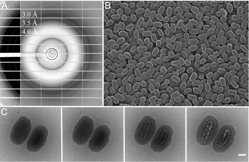
Fig. 1.
Granulovirus OBs contain a single virion surrounded by a crystalline protein layer that diffracts to high resolution. (A) Powder X-ray diffraction from a pellet of granulovirus OBs at 100 K (Materials and Methods). Protein diffraction rings extend to a resolution between 3 and 3.5 Å. The detector panels on the left with enhanced contrast show evidence of diffraction at even higher resolution. Resolution rings are shown at 4, 3.5, and 3 Å. (B) Freeze etch electron micrograph showing the uniform size distribution of the particles (Materials and Methods). (C) Cryo-EM. The sequence of four 20 e/Å2 exposures shows the effects of radiation damage on granulovirus OBs. The crystalline lattice is visible only in the first image and hydrogen gas bubbles produced by radiolysis eventually reveal the virion. (Scale bar, 100 nm.)

