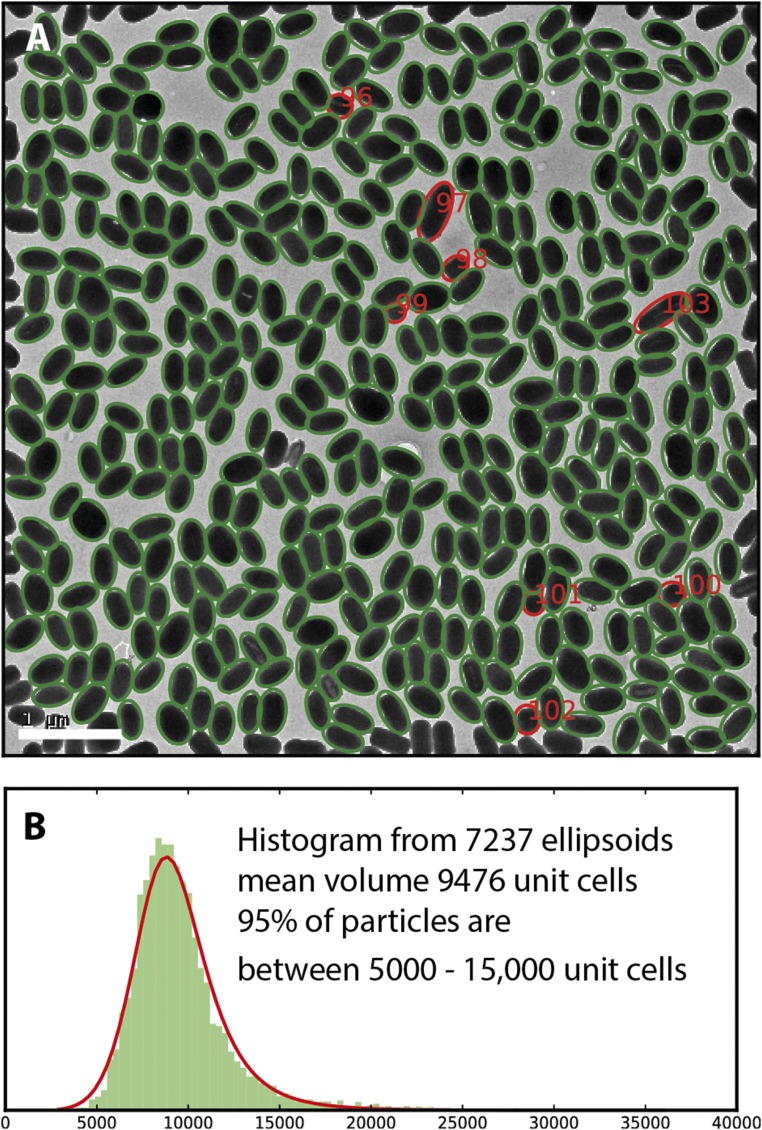
Fig. S1.
(A) Electron micrograph showing granulovirus occlusion bodies outlined by ellipses. The particles shown with red ellipses are outliers. Particles 97 and 103 are unusally large and the other red particles unusually small, probably because they are end-on views. (B) Histogram of volumes derived from 7,237 ellipsoids from particles in 16 similar images. The histogram has been fitted with a log-logistic distribution with mode ∼9,000 unit cells. According to this distribution, 2% of the particles are larger than 16,000 unit cells. (Scale bar, 1 µm.)

