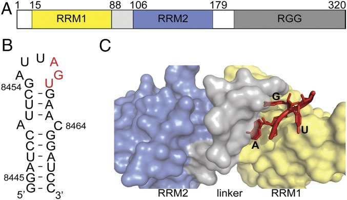
Fig. 1.
UP1 domain of hnRNP A1 binds specifically to 5′-AG-3′ through its nucleobase pocket. (A) Domain organization of full-length hnRNP A1. (B) Secondary structure of the HIV ESS3 (NL4-3 strain) stem loop wherein apical loop residues involved in UP1 binding are colored red. (C) Surface representation of UP1 bound to 5′-AGU-3′ (red). UP1 color coding is the same as in A.

