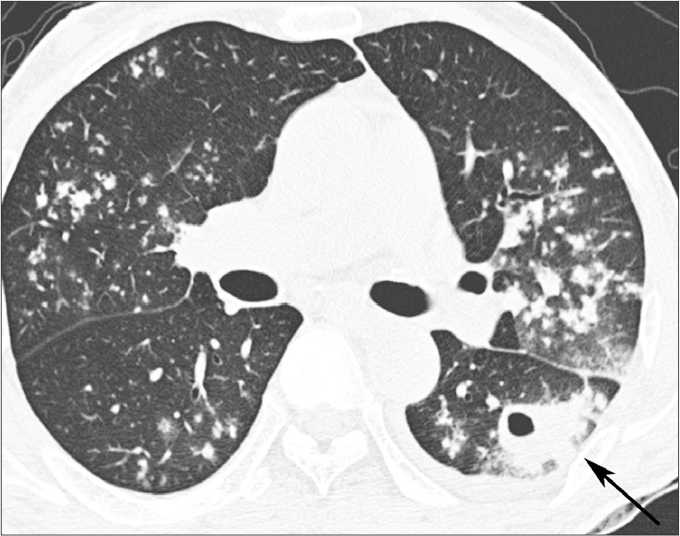
Figure 2.

A 59-year-old man with underlying DM and active pulmonary TB. High-resolution CT image shows bilateral lung involvement of active pulmonary TB. It shows multiple small nodules in bilateral upper lobes and superior segments of bilateral lower lobes. A cavitary mass (arrow) is located in the superior segment of the left lower lobe. A small amount of the left pleural effusion is noted.
