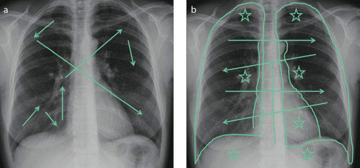
Figure 1. a, b.
Frontal view chest X-rays showing examples of different scanning pathways. A messy scanning pattern (a) in which the reader’s gaze jumps to different areas of the lungs without any method (arrows). An ordered scan path (b), which covers all the lung zones symmetrically (arrows). The “blind zones” (apices, hila, retro-cardiac and sub-diaphragmatic spaces) (stars) and the mediastinal lines and stripes (lines) should always be checked carefully.

