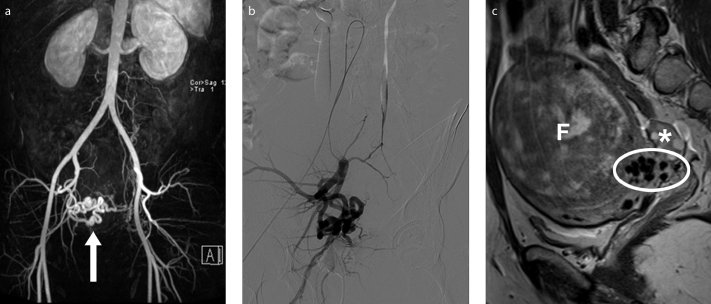
Figure 11. a–c.
Preprocedural MRI and MRA show an enlarged right uterine artery and a small caliber left uterine artery, in a 34-year-old-woman presenting for fibroid embolization. 3D reconstructed MRA image (a) depicts a prominent right uterine artery (arrow). DSA (digital subtraction angiography) image (b) obtained with selective right internal iliac artery catheterization correlates with the MRA findings. Right parasagittal T2-weighted image (c) show flow voids (ellipse) appearing as hypointense structures at the interface between the fibroid (F) and the myometrium, in relation with dilated arterial branches with fast flow, feeding the vascular plexus of the fibroid. Right ovary has unremarkable appearance (asterisk).

