
Figure 2.
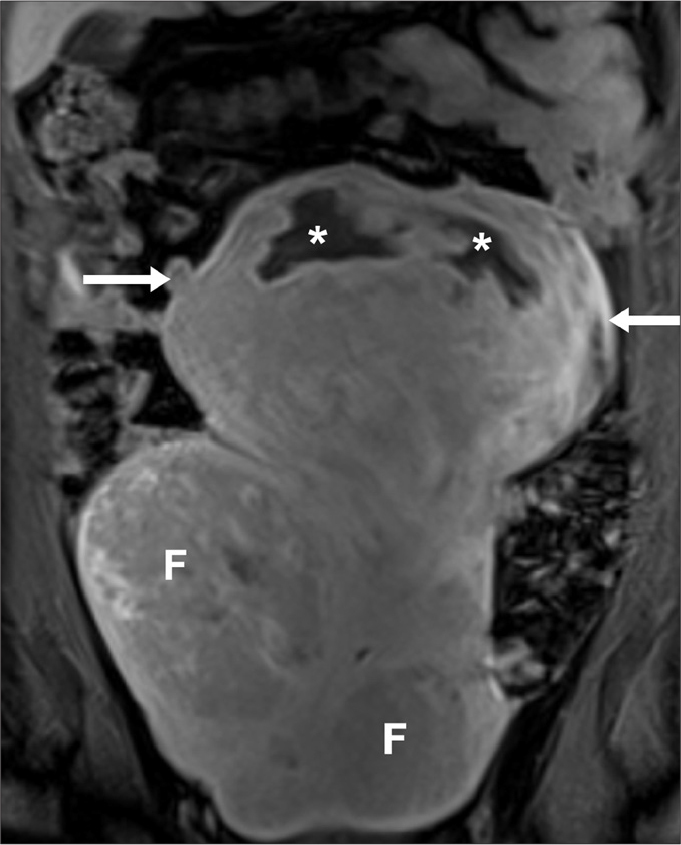
Suspicion of uterine leiomyosarcoma in a 31-year-old woman, presenting with epigastric pain and anorexia. The patient had undergone two previous uterine artery embolizations (UAEs). Coronal T2-weighted image obtained five years after the second UAE. The uterus is enlarged, extending to the upper abdomen. There is a bulky, heterogeneous mass (arrows) arising from the uterine fundus, with internal necrotic/cystic areas (asterisks). There are background uterine fibroids (F). Given the size and internal necrotic/cystic areas within the fundal lesion, the possibility of a leiomyosarcoma was raised. There was no evidence of metastatic disease. The patient underwent hysterectomy and histology revealed a fibroid.