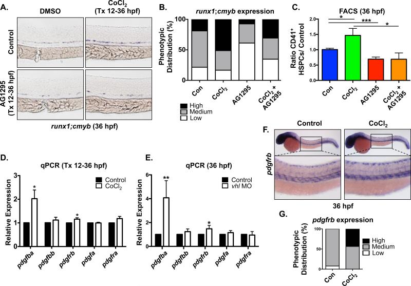
Figure 1. Hif1α stabilization enhances HSPC production via PDGF activity.
(A) Embryonic exposure to the pan-PDGFR inhibitor AG1295 (10μM) (during HSC formation (12-36hpf) decreased runx1;cmyb WISH expression in the AGM and blocked the increase in runx1;cmyb expression due to exposure to Hif1α agonist (CoCl2, 500μM). Tx = treatment.
(B) Qualitative phenotypic distribution of embryos from panel 1A scored with low, medium or high runx1;cmyb expression in the AGM (n≥20 condition × 3 replicate clutches).
(C) FACS analysis confirmed that chemical stabilization of Hif1α enhanced HSCs, while inhibition of PDGFR decreased CD41+gata1− HSPCs at 36hpf in the presence of CoCl2 (*p<0.05, ***p<0.001, one-tailed t-test, n≥13 replicates/condition).
(D) RT-qPCR analysis showed that pdgfba and pdgfrb were significantly upregulated over baseline in CoCl2 treated embryos, whereas related family members were not affected (Tx 12-36hpf) (*p<0.05, two-tailed t-test, n≥3).
(E) In vhl MO injected embryos RT-qPCR analysis likewise indicated that only pdgfba and pdgfrb were significantly upregulated over matched controls (Tx 12-36hpf) (*p<0.05, **p<0.01, two-tailed t-test, n≥4).
(F) WISH analysis of wild-type embryos at 36hpf indicated pdgfrb expression throughout the trunk of the embryo, including enrichment in the region of the VDA; pdgfrb expression was notably increased by CoCl2 exposure.
(G) Qualitative phenotypic distribution of embryos from panel 1F scored with low, medium or high pdgfrb expression in the AGM (n≥20 condition × 2 replicate clutches).

