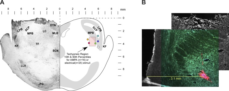
Fig. 3.
A: transverse section of rostral pons 1–2 mm caudal to the caudal pole of the inferior colliculus shows the locations where AMPA microinjections or electrical stimuli induced maximal tachypneic responses. Pink shaded box indicates the 10th and 90th percentiles of the stereotaxic coordinates of the Te-sensitive region. Filled circles indicate locations of fluorescent beads marking the location of the maximal tachypneic responses (n = 6 dogs). B: example showing fluorescent beads (red), medial to the superior cerebellar peduncle, where AMPA microinjections produced a marked tachypneic response. The image is a composite montage of 3 images to provide a more detailed perspective of the lateral and dorsal aspects of the transverse slice. DTN, dorsal tegmental nucleus; KF, Kölliker-Fuse nucleus; LC, locus coeruleus; LLV, lateral lemniscus ventral nucleus; LPB, lateral parabrachial nucleus; MLB, medial longitudinal bundle; MPB, medial parabrachial nucleus; PG, pontine gray; SCN, superior central nucleus; SCP, superior cerebellar peduncle; TF, tegmental field.

