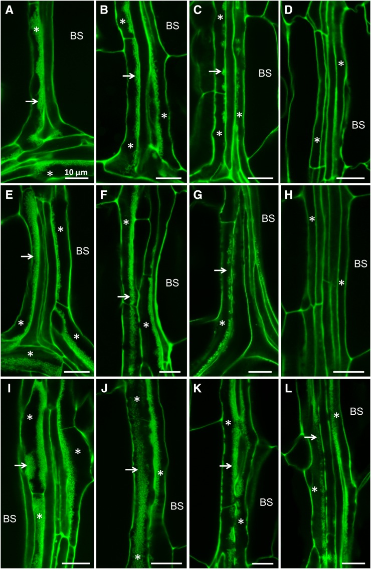
Figure 2.
Confocal imaging of wall ingrowth deposition in PP TCs in mature leaves of different ecotypes. A to D, Col-0. E to H, Ws-2. I to L, Ler-0. A, E, and I, Confocal images of veins from juvenile leaf 1 of Col-0 (A), Ws-2 (E), and Ler-0 (I) plants showing Class V PP TCs characterized by massive deposition of wall ingrowths (arrows) occupying a large volume of each PP TC (asterisks). B, F, and J, Confocal images of veins from transition leaves, namely leaf 6 of Col-0 (B), leaf 4 of Ws-2 (F), and leaf 6 of Ler-0 (J) plants, showing Class IV PP TCs with extensive deposition of wall ingrowths (arrows). C, D, G, H, K and L, Confocal images of veins from adult leaves, namely leaf 11 of Col-0 (C and D), leaf 6 of Ws-2 (G and H), and leaf 7 of Ler-0 (K and L). Adult leaves of Col-0 and Ws-2 have Class III PP TCs (asterisks) with clusters of wall ingrowths (arrows) at the leaf tip (C and G) and have PP cells (asterisks) without wall ingrowths at the leaf base (D and H). Class IV PP TCs are seen at the leaf tip (K) and Class III PP TCs at the leaf base (L) in adult leaf 7 of Ler-0. Scale bar = 10 μm for all images. BS, bundle sheath.

