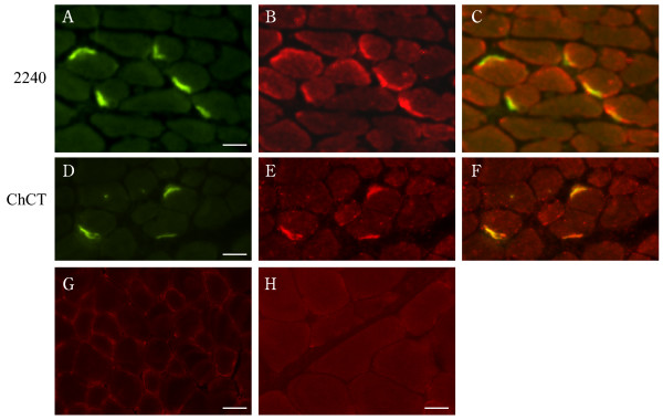
Figure 3.
Immunolocalization of CAR to the neuromuscular junction in mouse skeletal muscle. Frozen sections of mouse muscle were incubated with Alexa Fluor-conjugated α-bungarotoxin and antibodies to CAR as described in Materials and Methods. Panels A and D show α-bungarotoxin staining of acetylcholine receptors at neuromuscular junctions (green). Panel B shows immunofluorescent staining of the section in panel A with a polyclonal antibody (ab 2240) to the extracellular domain of CAR (red). Panel E shows immunofluorescent staining of the section in panel D with a chicken polyclonal antibody (ChCT) directed against a C-terminal portion of CAR conserved in both CAR isoforms (red). Panel C is a merge of panels A and B. Panel F is a merge of panels D and E. These merges [C, F] show that CAR colocalizes with α-bungarotoxin at murine neuromuscular junctions (in yellow). Sections incubated with the secondary antibodies alone did not give any signal – anti-rabbit IgG [G], anti-chicken IgY [H]. Bar = 35 μm

