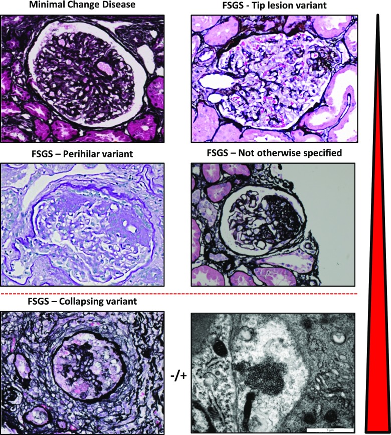
Figure 1.
Histopathology of minimal change disease and focal segmental glomerulosclerosis. Minimal change disease shows patent glomeruli in the absence of tubulointersitial scarring (silver stain, ×40). The tip lesion represents a focal adhesion of the glomerular tuft to Bowman’s capsule near the proximal tubule takeoff (silver stain, ×400). The most common forms of FSGS seen in adaptive FSGS and across all etiologies of FSGS are the perihilar variant (periodic acid–Schiff stain, ×40) and not otherwise specified pattern (silver stain, ×400). The most distinctive variant is the collapsing variant (collapsing glomerulopathy; silver stain, ×40). A specific instance of collapsing variant can be appreciated in the setting of endothelial tubuloreticular inclusions seen on ultrastructural analysis. These may be observed in high IFN states, including viral infection and exogenous IFN. The red arrowhead indicates the relative response to therapy and propensity of progression of these various forms, with minimal change disease and tip lesion being most responsive and least progressive and collapsing glomerulopathy being most therapy resistant and rapidly progressing.

