Figure 8.

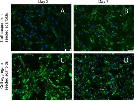
Confocal microscopy images of cardiac α‐actin (a marker of cardiac myocytes) and DAPI (cell nucleus) stained sections of cell suspension and cell aggregate seeded PUU scaffolds after 3 and 7 days in culture. The scale bar represents 25 μm.

Confocal microscopy images of cardiac α‐actin (a marker of cardiac myocytes) and DAPI (cell nucleus) stained sections of cell suspension and cell aggregate seeded PUU scaffolds after 3 and 7 days in culture. The scale bar represents 25 μm.