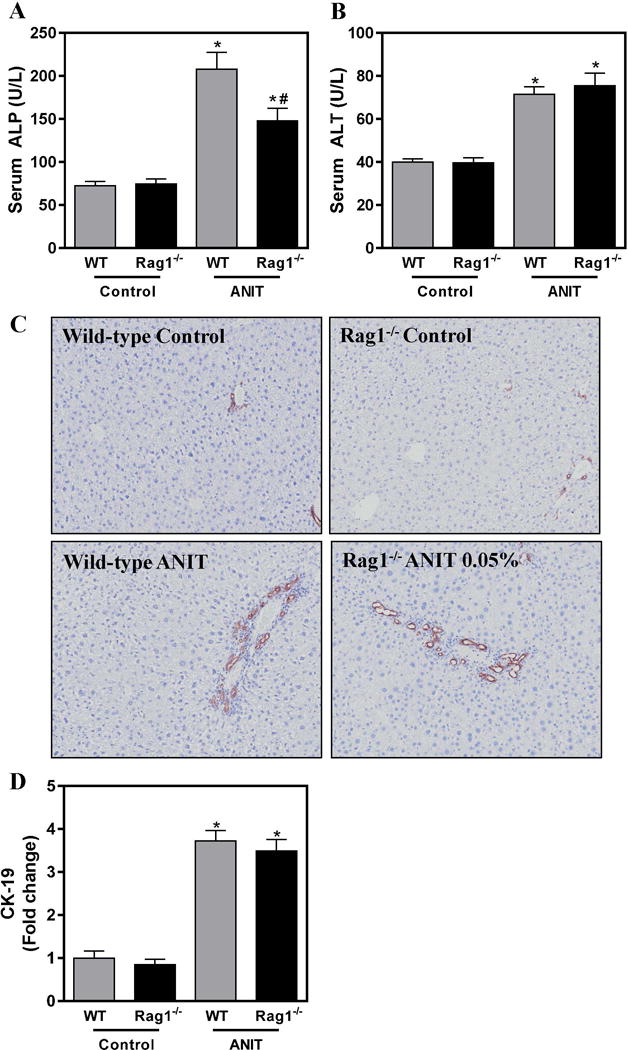
Figure 2. Lymphocytes contribute to biliary injury but not hyperplasia in ANIT-exposed mice.

Wild-type (WT) and RAG1−/− mice were fed standard control rodent chow or diet containing 0.05% ANIT for 4 weeks. (A) Serum ALP and (B) Serum ALT activity were determined as described in Materials and Methods. (C) Representative photomicrographs (100×) show liver sections stained for CK-19 (brown). (D) CK-19 staining was quantified as described in Materials and Methods. Data are expressed as mean + SEM; n=5–10 mice per group. *p<0.05 vs. control diet. #p<0.05 vs. ANIT-exposed WT mice.
