Figure 1.
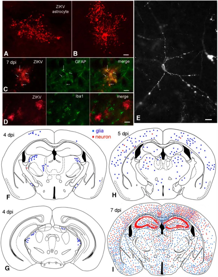
ZIKV enters brain after intraperitoneal inoculation. A, B, Confocal scanning microscope images of ZIKV-infected astrocytes. Scale bar, 8 μm. C, ZIKV-infected glial cell (red) contains GFAP immunoreactivity (green). The ZIKV immunoreactivity is found out to the tips of the glial processes, whereas the GFAP is confined more to the shaft of primary and secondary processes. Scale bar, 10 μm. D, No colocalization of ZIKV and Iba1 (a microglia marker) was detected. Scale bar, 12 μm. E, ZIKV-infected neuron with punctate immunoreactivity at 7 dpi after P0 inoculation. Scale bar, 15 μm. F, G, At 4 dpi after intraperitoneal inoculation, most infected cells are glia (blue dots); only rare neurons (red dots) are infected. G, More caudal midbrain region of the same mouse as in F. H, More cells, particularly astrocytes, are infected at 5 dpi. I, At 7 dpi after intraperitoneal inoculation, ZIKV has spread throughout the brain. At this stage of development, ZIKV infects glia (blue) and neurons (red) with little preference for brain regions, with the exception in this case of strong hippocampal neuron infection, including the dentate gyrus, CA3, and CA1.

