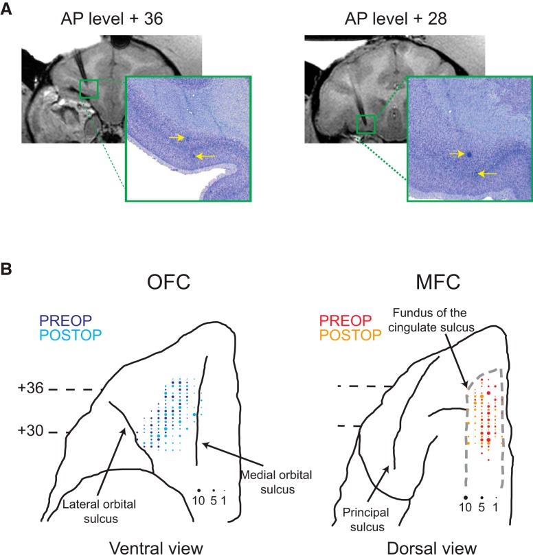
Figure 2.
Recording locations. A, Coronal T1-weighted MRI of electrode recording locations (left) and photomicrographs of Nissl-stained sections with corresponding marking lesions (right) at two different anterior–posterior levels (+36 and +28 mm anterior to the interaural plane) within OFC of Monkey V. Yellow arrows denote the location of marking lesions. B, Recording locations in OFC and MFC plotted on ventral (OFC) and dorsal (MFC) views of the frontal lobe from a standard macaque brain. Larger symbols represent increasing numbers of neurons recorded at that location. Darker colors (OFC: blue; MFC: red) represent preoperative recording locations, whereas lighter colors (OFC: cyan; MFC: orange) represent postoperative recording locations.

