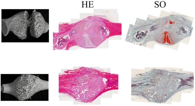
Fig 7. Microscopic images of fracture callus stained with hematoxylin and eosin (H&E) and safranin-O.
Representative images of non-union ribs are in the upper row and those of union in the lower row: micro-CT images (left column), histological images stained with H&E (middle column) and histological images stained with safranin-O (right column). Magnification = 40X, scale bar = 1 mm.

