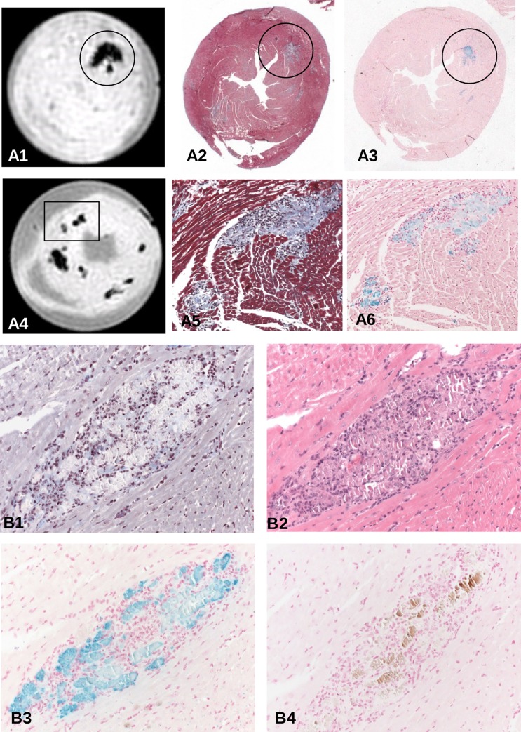
Fig 2.
(A) Spatial correlation of T2*w images of hearts 4 (A4-6, 9 days p.i.) and 2 (A1-3, 14 days p.i.) of Fig 1 with Masson's trichrome and Prussian blue-stained lesions. Iron deposits as stained by Prussian blue can be attributed to inflammatory lesions, mainly to affected cardiomyocytes at any time of acute and subacute myocarditis (A2, A3, x6; A5, A6 reflect area covering insert of A4, x200). (B) Dissection of an individual cardiac lesion by different histopathological stainings in consecutive tissue sections from a ABY/SnJ mouse 14 days p.i. (B1, Masson's trichrome, B2, HE, B3 Prussian blue, B4 von Kossa stain). Note, that the virus-induced damaged myocytes demonstrated in B1 and B2 spatially correspond well to iron deposits in myocytes (B3) whereas calcification (brown) occurs only in a part of the affected myocytes (B4) (x200).

