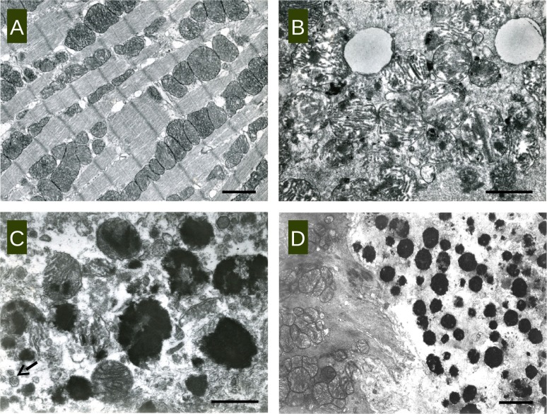
Fig 3.
Transmission electron microscopic image of representative heart tissue samples of ABY/SnJ mice (A) 0 days p.i. (uninfected control), (B) 6 days p.i., (C) 9 days p.i. and (D) 28 days p.i. Deposits of electro-dense material in mitochondria in association with structural disturbances of mitochondria and the cytoplasm of myocytes are observed at any stage of infection. (C arrow: virions; A, bar: 2 μm, B bar:1 μm, C bar:1,4 μm, D bar:2,5 μm).

