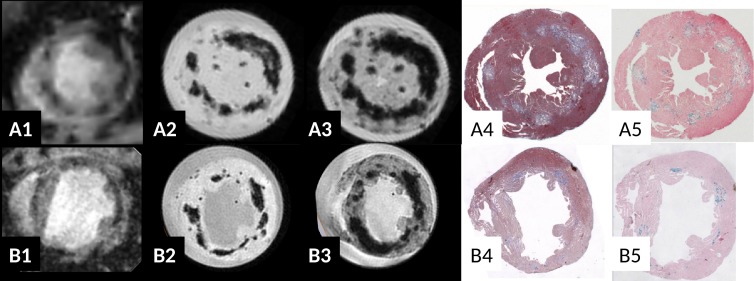
Fig 4. Correlation of T2* w contrast in vivo (A1, B1: TE = 2.6ms) and ex vivo (A2, B2 TE = 2.6/6ms) in chronic myocarditis in ABY/SnJ mice (8 weeks p.i., row A) and SWR/J mice (8 months p.i., row B).
The location of the T2* hypointense regions correlates very well with virus induced lesions as observed in Masson's trichrome staining (A4, B4) and Prussian blue staining (iron deposits, A5, B5). An increase in T2* blooming effect in ex vivo images is found at longer echo times. Note severe dilation of the left ventricle 8 months p.i. indicating DCM and heart failure in the SWR/J mouse 8 months p.i. (B).

