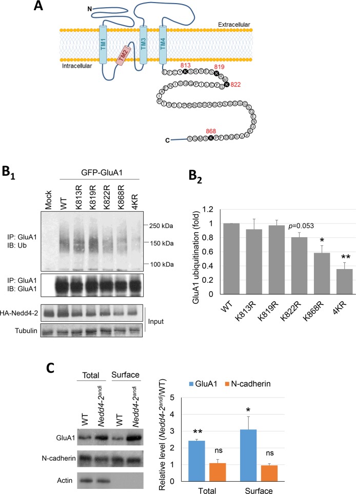
Fig 5. Nedd4-2 mediates surface expression of GluA1.
(A) Illustration of GluA1 on the cell membrane and the 4 lysine residues potentially ubiquitinated by Nedd4-2 at the C-terminus of GluA1. (B1) Western blots of Ubiquitin (Ub) or GluA1 after immunoprecipitation using anti-GluA1 antibody from HEK cells transfected with WT HA-Nedd4-2 along with WT GluA1 or mutant GluA1s (K813R, K819R, K822R, K868R and 4KR) for 48 hours. Quantification of ubiquitinated GluA1 by the area of smear from 100–250 kDa is shown on the right (B2) (n = 4, one-way ANOVA with post-hoc Tukey test). (C) Western blots of GluA1, N-cadherin, and Actin from WT or Nedd4-2andi cortical neuron cultures. Proteins from total lysate or after surface biotinylation were as indicated (n = 3, one-sample t-test was performed after normalization to WT groups). For all experiments, data are represented as mean ± SEM with *p<0.05, **p<0.01.

